皮膚病理組織学は、皮膚科学の中でも最も重要な位置を占める分野の一つであることは言うまでもありません。
しかしながら、最近では米国のように皮膚病理診断を自ら行わず、病理医/皮膚病理医に丸投げするという風潮が徐々に頭をもたげ、
また研究対象としての皮膚病理組織学を志す若い皮膚科医が激減してきております。
ここで再び、皮膚病理組織学の重要性を認識していただくために、
日本皮膚科学会理事長ならびに世界の著名な皮膚病理組織学者にお願いし、熱きメッセージを届けていただきました。
是非ご一読下さい。
日本皮膚科学会理事長からのメッセージ
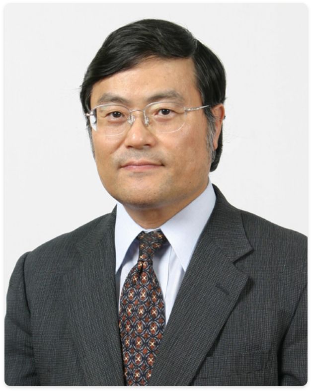
Shinji Shimada, MD
島田 眞路
日本皮膚科学会理事長
山梨大学皮膚科教授
「若き皮膚科医へのメッセージ」
皮膚病理学は皮膚科臨床医にとって最重要分野の一つです。皮膚の病理を知ることなく臨床皮膚科を修得することは不可能なほどです。日本皮膚科学会専門医試験でも皮膚病理学の問題は必ず出題され、しかも重要な位置を占めているのはそのためなのです。
東京大学では「組織デモ」と称する症例検討会があり、週一回各症例の病理組織学の発表・討論が行われていました。担当になった場合は学会発表並みに緊張したものです。思い返せばこれが私の皮膚科臨床の基礎になっているものと考えます。
米国留学時、皮膚病理専門医の存在を知りました。臨床カンファランスでは、彼らが活躍します。幅広い知識を有し、臨床なしでもHE染色標本だけでどんどん確実に診断して行く姿を見て、大変感銘を受けました。なかでも故アッカーマン教授の講演や検討会での発表・討論は圧巻でした。当時日本でもこのような専門医ができることを望んでいましたが、正式にはなかなか難しいようです。しかし本学会の中で、これに相当するような皮膚病理の専門家が育っていくことを期待しています。若手の方々には皮膚の病理が臨床皮膚科学の最重要分野であることを再認識してほしいと思っています。
世界の皮膚病理学者からのメッセージ
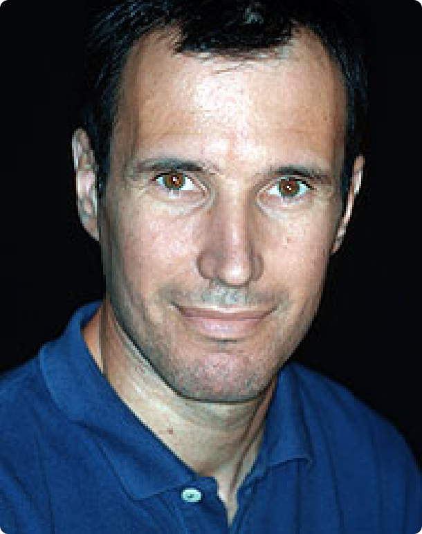
ボストン大学 Jag Bhawan 先生からのメッセージ

Jag Bhawan, MD
Professor of Dermatology and Pathology
Head, Dermatopathology Section
Vice Chairman, Department of Dermatology
Boston University School of Medicine
ボストン大学 Jag Bhawan 先生からのメッセージ
Dermatohistopathology is an important and integral part of learning as well as practicing dermatology. Its role in management of your patients can not be overemphasized. Not only is dermatohistopathology important in understanding basic disease process but it makes one a better dermatologist. As a student of dermatology, it is extremely important for you to learn the pathology of skin disorders, so that you are able to question your pathologist in any case of discrepancy between the clinical and your pathologist’s diagnosis. After all, you know your patient best and you must be able to correlate clinical and pathologic findings. Without solid knowledge in dermatohistopathology, this goal will be very difficult to accomplish. I can say this with great conviction as I am not only a trained dermatologist but a trained pathologist as well. Another dividend of dermatopathology is the ability for dermatopathologist to describe new entities or able to find pathobiology of disease process. For example, I had the fortune to describe intravascular lymphoma which in the past was mistakenly thought to be malignant tumor of endothelial cells (known previously angioendotheliomatosis proliferans systemisata). I would therefore encourage the younger dermatologits who are finishing up their training in dermatology to pursue dermatopathology as post graduate training for an exciting career. To underscore the importance of dermatopathology , we have 56 training programs in dermatopathology in USA which is highly competitive. We also offer dermatopathology to international trainees.
グラーツ大学 Lorenzo Cerroni 先生からのメッセージ
Lorenzo Cerroni, MD
Department of Dermatology
Medical University of Graz
Graz, Austria
グラーツ大学 Lorenzo Cerroni 先生からのメッセージ
Dermatopathology consists of macroscopic and microscopic skin pathology, and it represents the most important diagnostic test in Dermatology. It is a subspecialty of both Dermatology and Pathology, requiring clinical as well as pathological skills in order to be practiced at the best. It is particularly important that Dermatologists continue their long tradition in Dermatopathology (including famous persons such as Unna, Kaposi, Lever, Ackerman and Kerl among many others), as the knowledge of clinical Dermatology is a fundamental pre-requisite for a high level Dermatopathology. The future of Dermatopathology is bound to the experience, qualification, and knowledge of those doing it and on the passion and commitment of those caring for it.
Many of those practicing Dermatopathology in Japan are friends who have spent variable periods of time at my Institution, the Dermatopathology Unit of the Department of Dermatology, Medical University of Graz, Austria. It is thus my greatest pleasure to greet the Japanese Dermatopathologists from the pages of their web site!
ソウル大学 Kwang Hyun Cho 先生からのメッセージ
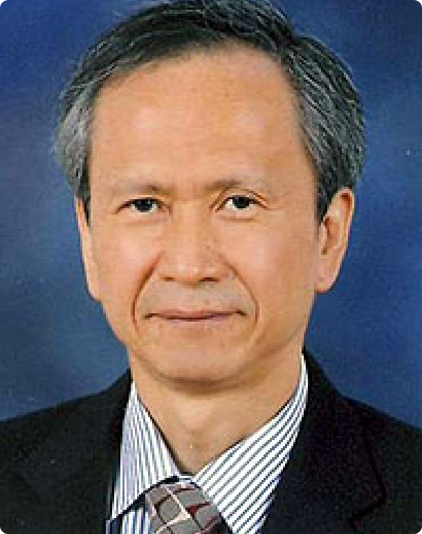
Kwang Hyun Cho, M.D.
Professor
Department of Dermatology
Seoul National
ソウル大学 Kwang Hyun Cho 先生からのメッセージ
To young dermatologists,
There are four traditional methods for clinicians to make a diagnosis- inspection, auscultation, percussion, and palpation. Among them, “inspection” is used to closely observe lesions and the most important way to make a diagnosis in the dermatologic field. Human trials to directly see the patient’s lesion achieved development of various medical instruments, including endoscopy and imaging devices. For example, endoscopy makes us see the problem, get a diagnosis, and treat the lesion. In this aspect, we, dermatologists have had a lot of advantages because we can personally see the lesion early on.
However, gross inspection alone is not sufficient for us to make a proper diagnosis. Just as gross pathology or clinical finding is important to pathologists, micropathology is to dermatologists. Gross inspection and histopathologic findings are complementary to each other, which can lead us to correct diagnosis. Therefore, histopathology is a basic knowledge for every dermatologist should know. The cornerstone of dermatology is to understand the normal skin structures and pathologic changes of various skin diseases. In every field of dermatology, whether it is basic, clinical, or aesthetic, you should start your step with studying dermatopathology.
元日本皮膚病理組織学会理事長
小野友道 先生からのメッセージ
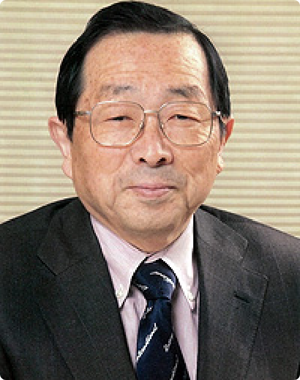
Tomomichi Ono
小野友道
熊本大学名誉教授
熊本保健科学大学学長
元日本皮膚病理組織学会理事長
小野友道 先生からのメッセージ
3Dを駆使している皮膚科医
皮膚科医以外に病理組織診断が出来る医師は極めて少ない。ほとんどが病理医に任せきりである。すくなくとも小生の現役時代はそうであった。皮膚科医のこの得意芸をわれわれはきっと大事にしてゆかねばならない。
今日、医学医療における診断技術はすばらしい発展を遂げている。CT然りである。それは人体のいろいろ断面を見事に表出させ、医師をして的確な診断に必要な情報を提供してくれる。そして得られたデーターから、例えば見事な3次元象を見せてくれる。
とまれ、皮膚疾患の臨床象と、その病理組織切片との組み合わせは、CTが示してくれる情報と全く同じではないか。いや、触覚、聴覚、嗅覚をも駆使して臨床象を観察し、断面象である組織切片がセットで前にある。その断面象をいかようにも染め上げ有用な情報を引き出せるではないか。ようやくCTなどの器機が皮膚病診断に少し近づいてきたのである。
臨床象を鋭く観察し、その内に潜む病変を、表皮真皮に至るまで想定できるのは、ひとえに皮膚病理診断技術を鍛えている皮膚科医を於いてないのである。
臨床と病理の両者を知り、はじめて皮膚科医と言えるほど皮膚科医にとって皮膚病理診断は不可欠なものでるといえる。
若い皮膚科医の諸君のなかに病理診断を病理医にだけ依存して、自分自身で顕微鏡を眺めないものが居るとすれば、それは嘆かわしいことである。病理医に診断を求めることはもとより極めて重要で、われわれ皮膚科医には到底及ばぬ診断技術を持っているのは承知している。それを十分吸収して、さらに自分自身で顕微鏡を眺めながら、治療方針にまで考えを及ぼしてほしいのである。
太田正雄が太田母斑の組織象を描いている。ひょっとしたら画家に描かせた可能性もあるが、少なくとも横で緻密に教示しながらものにしたと思われるが、真皮のリボン状の色素細胞(今日で言うところのdermal melanocyte)とともに、被覆表皮基底層にも通常より多くのmelanocytesが描かれている。どれだけ丁寧に観察がなされたか。どれだけ臨床を思い浮かべて観察したか、凄いとしか言いようがない。